Download pdf
Case #31921377
Summary: Unilateral transient visual loss during coughing spell
History
Presentation: 41-year-old woman has had two episodes transient vision loss.
Inquiry: Bilateral vs Unilateral
nilateral, first episode was in the right eye and second episode was in the left eye
Inquiry: Duration
1-2 minutes per episode
Inquiry: Episode Characteristics
Complete black out of vision
Inquiry: Activity during the episode
Both episodes occurred during an intense coughing episode
Inquiry: Pattern of visual loss
She was able to cover each eye during one of the episodes to determine that it was her left eye during the first episode that went dark and it was the right eye during the second episode.
Inquiry: Other Symptoms
She recently has tried to stop smoking cigarettes and as a result, has been experiencing frequent coughing spells.
Inquiry: Current Medication
Medications include birth control pills and nicotine patches.
Exam Results
Ophthalmologic exam was normal with normal anterior segment and retina and optic nerve in each eye.
Ocular motility was normal and there was no relative afferent pupil defect.
Intraocular pressures were 13 mmHg OD and 14 mmHg OS.
Commentary
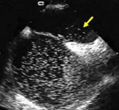
Because each episode occurred in different eyes, one is thinking of a more proximal source of emboli, which would be the heart and/or aorta. Because of the occurrence with coughing, one should consider a paradoxical embolus. Venous clot moving into the right chamber of the heart from the great veins returning blood to the heart and then shunting to the left chamber through an opening in the septum of the heart wall (patent foramen ovale). During coughing the venous pressure rises abruptly forcing a small clot to pass through the opening connecting the two chambers of the heart. Normally, without the opening, such particles would be filtered out by the lungs before returning to the left chamber of the heart.
So in this case an echocardiogram is indicated and a “bubble” study (echocardiogram with contrast). Small bubbles that are echogenic are injected into the arm vein and as they pass into the right heart chamber they are seen with ultrasound. If there is a patent foramen ovale, then the bubbles pass directly from the right to the left chamber through the opening.